normal epiglottis thickness
Skip to main content. It can safely exclude the acute epiglottitis.
Learningradiology Epiglottitis
On CT imaging the Halloween sign describes an epiglottis of normal thickness.
. There was significant correlation between epiglottis thickness and body surface area r0533 weight 0517 height 0437 and body mass index 0372. The average Korean epiglottis thickness was measured to be 231-022 mm range. Epiglottis width of 55 mm or more was found to be 962 sensitive and 100 specific in diagnosing acute epiglottitis.
We believe that these data could serve as a reference in diagnosing and detecting abnormalities. 188 mm to 284 mm with greater thickness noted in men 241-021 mm when compared to women 221-018 mm p 0001. The epiglottis is a cartilaginous flap that extends in front and above the laryngeal inlet or more specifically the rima glottidis glottis.
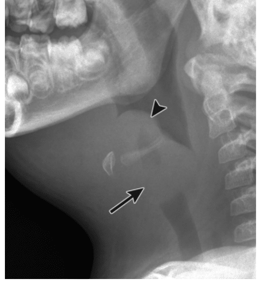
This 30-year-old man was wounded with a shotgun. However one pellet entered the mental vertex of his mandible and lodged in the anterolateral neck. Figure 4-11 Penetrating neck trauma.
The normal epiglottis is a thin crescent. We believe that this would facilitate diagnosing and determining the extent of cancerous invasion of the supraglottis and secondary invasion of the epiglottis. Wpr-198568 WPRIM ID.
The epiglottis in men was significantly thicker than that in women p. Journal Article OPEN ACCESS. The average thickness of epiglottis was 0236 cm and the standard deviation was 0020.
An epiglottic width greater than 8 mm or aryepiglottic fold width greater than 7 mm is suggestive of epiglottitis. During swallowing the epiglottis folds back over the larynx inlet thus prevents foods from entering the airway. No statistically significant difference was observed in thickness depending on side or age.
Gross anatomy The epiglottis projects posterosuperiorly from its stem-like base which is attached to the thyroid cartilage. The epiglottis is a single midline leaf-shaped fibrocartilaginous structure that forms part of the supraglottic larynx and defines the division of the hypopharynx from the larynx. No statistically significant difference was observed in thickness depending on side or age.
It should be noted that an omega epiglottis either a variant of normal or in the setting of laryngomalacia can result in a similar appearance and can be mistaken for epiglottitis. The epiglottis is usually 3-5. Positive and negative likelihood ratios were infinity and 004 respectively.
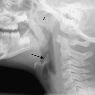
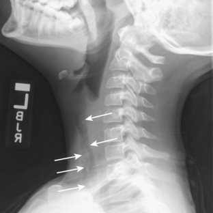
B c Sagittal b and axial c contrast-enhanced CT images in a 21-year-old man with epiglottitis show thickening of the epiglottis arrow in b and aryepiglottic folds arrows in c. The thickness at the median was larger than that bilaterally in all patients p. A statistically significant difference was observed in thickness depending on longitudinal height p.
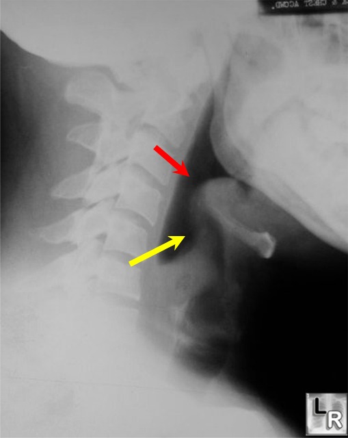
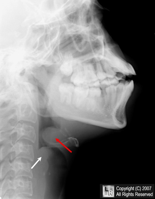
The edematous epiglottis appears enlarged and thumb-shaped Fig. Journal of the Korean Society of Emergency Medicine. The mean and median of EP2s measurement were slightly smaller than those of EP1 so were the lower and upper quartiles.
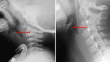
The thickness at the median was larger than that bilaterally in all patients p. Soft-tissue lateral neck radiograph reveals edema of epiglottis consistent with acute epiglottitis. See the image below.
To our knowledge this is the first study to investigate the thickness of the normal epiglottis on computed tomography CT in a Japanese po. The function of the epiglottis is to close the laryngeal inlet during swallowing and so to prevent the passage of food and liquid into the lungs aspiration. Halloween sign describes the CT appearance of a normal-thickness epiglottis Epiglottis A thin leaf-shaped cartilage that is covered with laryngeal mucosa and situated posterior to the root of the tongue and hyoid bone.
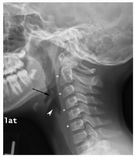
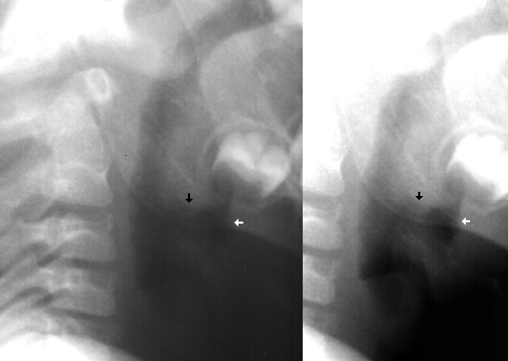
WPRIM ID. Aryepiglottic fold width of 59 mm or more was 923 sensitive and 808 specific. Normal epiglottis versus epiglottitis in two patients.
This retrospective study was based on a review of radiographic data in patient. Like the guttural pouches and pharynx the larynx is optimally imaged with a lateral projection of the throat using a soft-tissue exposure. The patient ducked his head at the moment of the blast and most of the shot impacted superficially in his scalp.
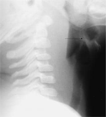
A Sagittal contrast-enhanced CT image in a 41-year-old man shows a normal epiglottis arrow measuring 23 mm in thickness. Download Citation Sonographic Measurement of the Epiglottis in Normal Chinese Adults Objectives 1 To assess the normal range of thickness of the epiglottis by means of ultrasound measurement. Furthermore CT imaging can help to diagnose other conditions such as peritonsillar abscess or retropharyngeal abscess which.
Evaluation of Normal Epiglottis on Computed Tomography with Special Attention to Thickness. The focus was on determining the thickness of a normal epiglottis which could then serve as a reference in detecting abnormalities. This is why we cant and shouldnt try to talk.
The epiglottis is a leaf-shaped flap of cartilage located behind the tongue at the top of the larynx or voice box. Lateral soft tissue radiograph of the neck demonstrates thickening of the epiglottis and aryepiglottic folds referred to as the thumb sign. The thickness of the normal epiglottis was established at each level.
An estimate of the thickness of the epiglottis of the normal population. There was an outlier in EP2s measurement which corresponded to a subject whose epiglottis. Adults with suspected epiglottitis and normal soft tissue radiographic films.
The Normal Epiglottis The radiographic appearance of the equine epiglottis is variable being subject to technical anatomic and physiologic influences. The main function of the. Artículo en Ko.
Sign In Create account.
Acute Epiglottitis Imaging

Epiglottitis Radiology Reference Article Radiopaedia Org
2

Ct Scans Of The Larynx Showing A Thickening Of Aryepiglottic Folds Download Scientific Diagram
Upper Airway Obstruction Radiology Key

Normal Anatomy Trachea And Bronchi Euroform Healthcare
Imaging Soft Tissues Of The Neck Radiology Key
Learning Radiology Epiglottitis

55 Year Old Man With Epiglottic Calcification Findings Axial A Download Scientific Diagram
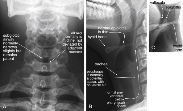
Airway Radiology Key

Epiglottitis Radiology Reference Article Radiopaedia Org
How Can We Rule Out Epiglottitis Don T Forget The Bubbles
2
Epiglottitis Teachmepaediatrics

Epiglottitis Radiology Reference Article Radiopaedia Org
Acute Epiglottitis Imaging
Airway Radiology Key

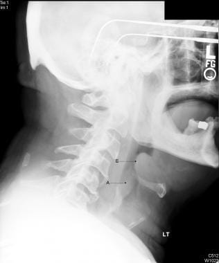
Normal Lateral Soft Tissue Radiograph Of The Neck In A Paediatric Download Scientific Diagram
Acute Epiglottitis Imaging


Comments
Post a Comment